
Dental X-ray and Microscope in Root Canal Therapy Examination
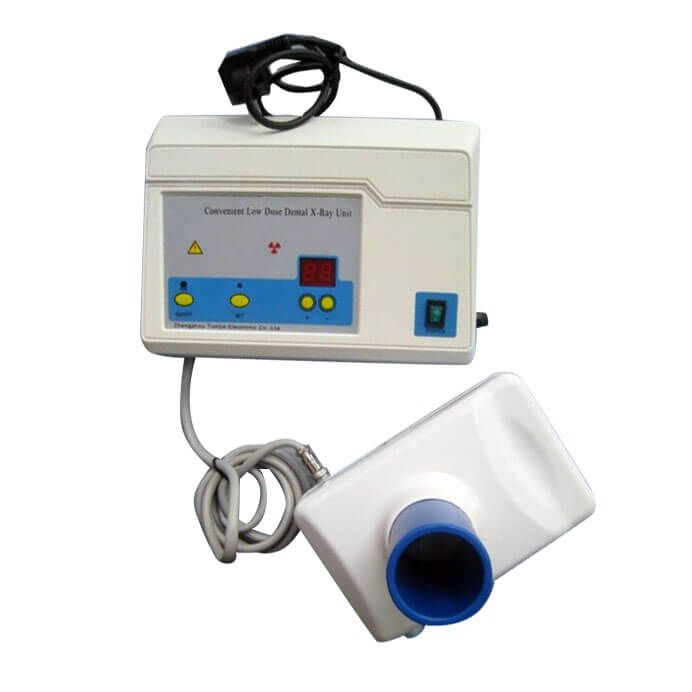
To put it simply, we want to see what happens to the teeth inside the bones and the teeth inside the teeth? So we invented a perspective machine, dental X-ray. It’s a bit like when we are at the airport, we use X-ray machine to “see” our luggage and check whether we have dangerous goods.
Dental X-ray: it can be roughly divided into single tooth X-ray pictures of two or three teeth, ring mouth X-ray pictures of the whole mouth, and other pictures such as wing biting film, temporomandibular joint X-ray, nasal sinus X-ray, and different angles of head used for correction, etc.
Dental cone beam CT: it can be simplified as an advanced version of dental X-ray. Through different principles and calculations, it can present 3D stereoscopic views of the mouth and teeth, as well as different sections and different axis terms, so that doctors can get more information.
Shooting time: in terms of dental treatment or root canal treatment, X-ray films are required before, during and after treatment.
Objective: the location of abscess, tooth structure, root canal information: length, number, location of root canal treatment, periodontal information, bone structure beside teeth, paranasal sinus, mandibular nerve canal, temporomandibular joint, etc.
Microscope assisted examination
Our naked eyes have a limit. We can distinguish the thickness of 0.1mm-0.2mm at least, almost the thickness of false eyelashes, but we can’t recognize the finer ones. The average resolution of dental microscope is 6 μ m, that is 0.006mm, which is more subtle than that of our naked eyes.
The dental microscope can help doctors to achieve better minimally invasive operation, which has been widely used in dentistry.
The timing of root canal therapy is as follows:
- Special examination (such as tooth mark, root canal perforation, root fracture, etc.).
- Search for additional nerve canal, such as the second nerve root canal near the buccal side of the maxillary molar.
- Special root canal (such as type C nerve canal).
- Remove obstruction.
- Deal with small or calcified neural tube.
- Repair and root tip operation.
- The place where the apical opening does not close the teeth (such as vital pulp treatment, vital pulp preservation, etc.).
- The whole procedure of root canal therapy was used.
Precautions for toothache
Clinical often found that many patients before, during and after treatment, it’s very easy to feel like trying to see this tooth by themselves? Is it good? Does it hurt? So I think of knocking, pressing, shaking, biting and sticking my tongue to my teeth. I want to “play and test myself” or see how these conditions become.
These tests should be done by the doctor. It is not recommended for the patient to try them on their own, which may aggravate your symptoms. Because the teeth that are supposed to rest and repair have been “knocked and bit” by you all the time, but without rest, it is not easy to “get well”.


